Volume 32
-
No. 12 December 2019
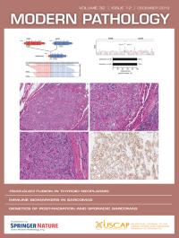
The cover shows representative micrographs and the recurrent PAX8-GLIS3 fusion gene of hyalinizing trabecular tumors of the thyroid. For more information, see the paper by Marchiò et al p 1734, this issue.
-
No. 11 November 2019
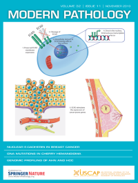
The cover shows a proposed model of nuclear translocation of E-cadherin in lobular breast cancer. For more information, see the paper by Lobo et al, p 1574, this issue.
-
No. 10 October 2019
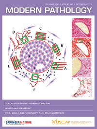
The cover shows the different parameters assessed to evaluate the collagen characteristics surrounding breast ductal carcinoma in situ. For more information, see the paper by Toss et al, p 1473, this issue.
-
No. 9 September 2019
The cover shows magnetic resonance imaging, histology and immunohistochemistry of high-grade infiltrating glioma of the spinal cord. For more information, see the paper by Alvi et al, p 1236, this issue.
-
No. 8 August 2019
The paper by Ma et al (p. 1217) shows that SATB2 and CDX2 are prognostic biomarkers in DNA mismatch repair protein-deficient colon cancer. The cover shows examples of colonic adenocarcinomas with loss of SATB2 and CDX2 expression by immunohistochemistry.
-
No. 7 July 2019
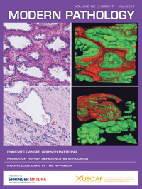
The paper by Verhoef et al, this issue, describes the two major architectural subgroups of prostate cancer growth patterns. The cover shows hematoxylin & eosin slides and three-dimensional renderings of the peripheral zone. For more information, see the paper on p. 1035.
-
No. 6 June 2019
On the cover: Whole lung microCT imaging with showing airways in obstructive and restrictive graft vs. host disease lungs. For more information, see the paper by Verleden et al, this issue, p 825.
-
No. 5 May 2019
Zhu et al validated and implemented a clinical molecular diagnostic assay, MSK-Fusion Solid, for detection of gene fusions in solid tumors including sarcomas. The cover shows a Circos plot of the 78 gene fusions identified from all the soft tissue and bone tumors submitted the assay, and representative sarcomas. For more information, see the paper on p. 609, in this issue.
-
No. 4 April 2019
The cover shows a selection of inflammatory myopathies and necrotizing myopathies using various staining techniques. For more information, see the paper by Cai et al, p. 462, in this issue.
-
No. 3 March 2019
Trimethylation of lysine 27 of histone H3 (H3K27me3) is a hallmark mechanism of transcriptional silencing. The cover shows examples of H3K27me3-deficient chondrosarcomas. For more information, see the paper by Makise et al, p435 this issue.
-
No. 2 February 2019
In this issue, The cover shows pathologic and radiographic findings of intraosseous synovial sarcoma. There is a fleshy intramedullary lesion in the metaphysis without a significant soft tissue component. Conventional lateral radiograph and axial CT showed a subtle mixed lytic-sclerotic lesion in the tibial metaphysis; the circumscribed oval lytic lesion on the conventional radiograph is a biopsy cavity. For more information, see the paper by Horvai et al, p231, this issue.
-
No. 1 January 2019
In this issue, the paper by Lin et al describes how hTERT promoter mutations in chondrosarcomas associate with progression and disease-related mortality. The cover shows a single case of chondrosarcoma with mixed histology (see December 2018 issue, p1834).