Volume 34
-
No. 12 December 2021
In this issue, the paper by Brück et al (p 2229) discusses spatial immunoprofiling of the intratumoral and peritumoral tissue of renal cell carcinoma patients. The cover shows images of immune topographies and corresponding cell detection and classification results, with lymphocytes in red and non-lymphocytes in yellow.
-
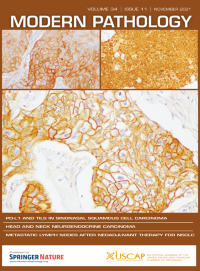
No. 11 November 2021
The paper by Hongo et al, this issue (p 1966), analyzes PD-L1 expression, tumor-infiltrating lymphocytes, mismatch repair deficiency, EGFR alteration and HPV infection in sinonasal squamous cell carcinoma. The cover shows immunohistochemical staining for PD-L1, EGFR, and p16 in patient samples.
-
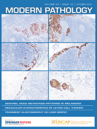
No. 10 October 2021
Cover caption: The cover shows microanatomic melanoma metastasis patterns within sentinel nodes. For more information, see the paper by Kretschmer et al, p 1839, this issue
-
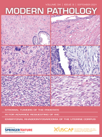
No. 9 September 2021
The cover shows the morphologic spectrum of tumors of purported specialized prostatic stromal origin with gene rearrangements and TP53 mutations. For more information, see the paper by Acosta et al, p 1763, this issue.
-
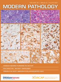
No. 8 August 2021
The cover shows representative histological images of HMGA2-NCOR2 fusion-positive giant cell tumors. For more information, see the paper by Agaimy et al p 1507, this issue.
-
No. 7 July 2021
A retrospective single-center study was conducted on patients with non-small cell lung cancer resected after neoadjuvant treatment. The cover shows representative H&E slides for estimation of the residual tumor. Light blue: demarcation of tumor tissue. Yellow: demarcation of necrosis. Green: demarcation of cholesterol crystals. Red: demarcation of thickened/hyalinized vessels. Black: demarcation between tumor/necrosis. For more information, see the paper by Zens et al, this issue, p 1333.
-
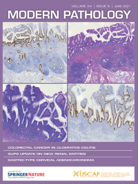
No. 6 June 2021
The cover shows histology and TP53 immunohistochemistry of ulcerative colitis-associated colorectal carcinoma. For more information, see the paper by Hirsch et al, p 1153, this issue.
-
No. 5 May 2021
The cover shows a model of synchronous endometrial and ovarian cancer development in Lynch syndrome and sporadic settings. For moreinformation see the paper by Moukarzel et al, p 994, this issue.
-
No. 4 April 2021
The cover shows NEAT1-TFEB rearranged renal cell carcinoma. For more information, see the paper by Caliò et al, p 842, this issue.
-
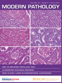
No. 3 March 2021
The cover shows the main pathological features of biphasic squamoid alveolar papillary renal cell carcinoma. For more information, see the paper by Denize et al, p 647, this issue.
-
No. 2 February 2021
The paper by Harmon et al (p 478, this issue) describes a high-throughput assessment of biomarkers in tissue microarrays using artificial intelligence. The authors show PTEN loss as a proof-of-principle in multi-center prostate cancer cohorts. The cover shows examples of true positive cases, correctly identified as having PTEN loss from internal and external testing cohorts.
-
No. 1 January 2021
The cover shows histological and immunohistochemical features of cervical carcinomas with serous-like papillary and micropapillary components. For more information see the paper by Wong et al, p 207, this issue.