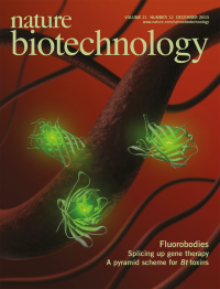
Volume 21
-
No. 12 December 2003
A representation of fluorobodies, 'sticky' fluorescent proteins that combine the binding specificity of antibodies with the fluorescence of green fluorescent protein, binding to a microtubule (see Zeytun et al. p. 1473). Graphic by Ken Eward, © BioGrafx.
-
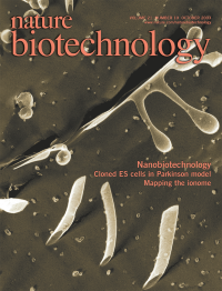
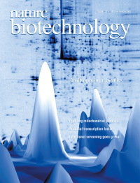
No. 11 November 2003
3D image of the mitochondrial compartment of a live yeast cell obtained through volume rendering of a superresolution 3D data stack recorded with a 4Pi microscope. Courtesy of H. Gugel, R. Storz (Leica Microsystems Heidelberg GmbH, Mannheim, Germany) and J. Bewersdorf, S. Jakobs, J. Engelhardt, S.W. Hell (MPI for Biophysical Chemistry, Göttingen, Germany). The 3D image is superimposed on a colored high resolution scanning electron micrograph of mitochondria and rough endoplasmic reticulum. © PhotoResearchers. Artwork rendered by Erin Boyle.
Focus
-
No. 10 October 2003
Quick-freeze/deep-etch transmission electron microscope (TEM) images of a solution of peptides that form peptide nanotubes. There are also vesicles budding off of a nanotube and micelles in the area. Peptide nanotubes may provide new biomaterials for materials science and biology research (see Zhang, p. 1171). Peptide nanotubes prepared by Sylvain Vauthey and Steve Santoso. Artwork rendered by Erin Boyle.
Focus
-
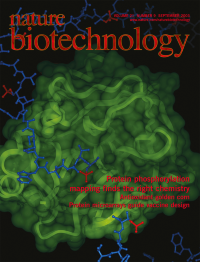
No. 9 September 2003
Trypsin cleaving a hypothetical protein at sites of phosphorylation, liberating a series of C-terminally phosphorylated peptides (see Knight et al. p 1047). The image was provided by Zachhary Knight at University of California, San Francisco and was made using the graphics program InsightII, rendering the crystal structure of trypsin as a ribbon underneath a transparent Connelly surface. The substrate is rendered as ball and stick, the phosphate groups in red. Artwork rendered by Erin Boyle.
-
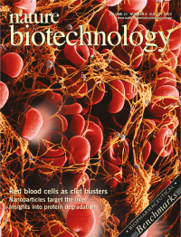
No. 8 August 2003
A representation of fibrinolysis at a clot by tPA-coupled red blood cells (see Murciano et al. p 891). Artwork by Erin Boyle. Image of blood clot © PhotoResearchers
-
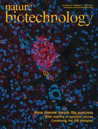
No. 7 July 2003
A cross section from a mouse pancreas reveals an emerging islet, with insulin stained red and nuclei stained blue. On p. 763, Bhatia and colleagues show that transplantation of bone marrow cells in a mouse model of diabetes induces pancreatic regeneration.
-
No. 6 June 2003
Visualization of the reporter β-galactosidase in various tissues of knockout mice reveals distinctive patterns of gene expression. On p. 822, Valenzuela et al. describe a high-throughput method for generating the mice.
-
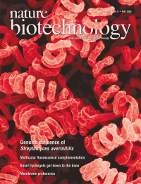
No. 5 May 2003
The genome of Streptomyces avermitilis may yield new antibiotics (see p. 526).
-
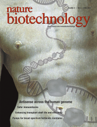
No. 4 April 2003
Molecular representation of transcription from a section of the human genome superimposed on Michaelangelo's David. (Image courtesy Rotman and colleagues, p. 379)
-
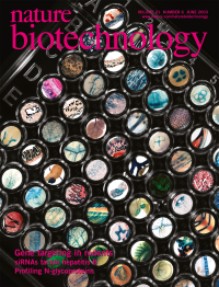
No. 3 March 2003
This issue focuses on the technological challenges in proteomics. (Image by Doug Huff, Arkitek Studios Inc., Seattle, WA. Used with permission of the Plasma Proteome Institute, Washington, D.C.).
Focus
-
No. 2 February 2003
Cover shows budding yeast superimposed on a steroid metabolic pathway. On p. 143, Dumas and colleagues demonstrate that rewiring of metabolic networks in yeast shows promise for biosynthesis of complex molecules of pharmaceutical interest from simple carbon sources. Yeast image © Photo Researchers.
-
No. 1 January 2003
Cover shows cocooned silkworms in a circular nest at a silk farm. On page 52, Yoshizato and coworkers report collagen production by transgenic silkworms. (Photo courtesy of Stephanie Colasanti/CORBIS).