Volume 17
-
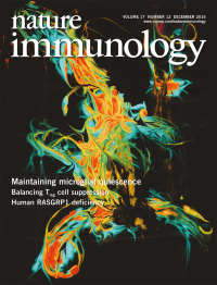
No. 12 December 2016
Microglia are resident macrophages in the central nervous system. Greter and colleagues (p 1397) demonstrate that the transcriptional regulator Sall1 is expressed specifically by microglia and is critical for microglial identity and homeostatic functions. The original image by Anne Buttgereit shows a three-dimensional reconstruction of a wild-type microglial cell stained with Iba-1. Artwork by Lewis Long.
-
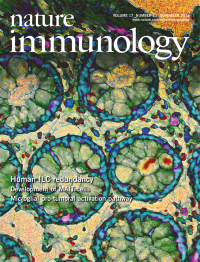
No. 11 November 2016
The contribution of innate lymphoid cells (ILCs) to immunity in natural conditions is unclear. By studying circulating and tissue-resident ILCs in patients with severe combined immune deficiency, Vély and colleagues (p 1291; News and Views by Le Gros et al. p 1237) show that ILCs are dispensable for immunological protection in the context of modern medicine and hygiene. The original image by Julie Bruneau shows scattered NKp46+ ILCs (brown) in the lamina propria and T cells (red) in the epithelium of a colonic biopsy. Artwork by Lewis Long.
-
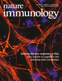
No. 10 October 2016
The immune system needs to tailor its responses according to the threat level. Lemaitre and colleagues (p 1150; News and Views by Tomé-Poderti and Saleh, p 1138) identify a novel version of the peptidoglycan receptor PGRP-LC in flies and demonstrate that this 'tunes' immune responses according to challenge with live (threatening) bacteria or dead (harmless) bacteria. The original image by Claudine Neyenis shows a section through fat-body cells expressing a mutant form of the receptor (green). Artwork by Lewis Long.
-
No. 9 September 2016
The contribution of stromal cells to the microenvironment of tumor-draining lymph nodes is poorly characterized. By comparative transcriptional analysis, Shields and colleagues (p 1118) find that tumors induce stromal reprogramming of key pathways that affect the structure and function of such lymph nodes. The original immunofluorescence image shows a resting lymph node with collagen-I-secreting stromal cells (blue), T cells (green) and B cells (red). Artwork by Lewis Long.
-
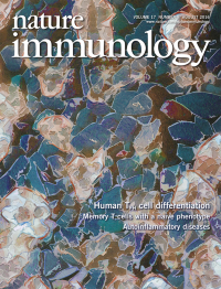
No. 8 August 2016
Knowledge about the signals that drive the differentiation of human follicular helper T cells is still very limited. Crotty and colleagues (p 976) demonstrated that the cytokine activin A, which is physiologically expressed in sites in which such differentiation is initiated, regulates multiple facets of the biology of these cells. The original confocal image by Zbigniew Mikulski is a Hoechst stain of cells in a human tonsil section. Artwork by Lewis Long.
-
No. 7 July 2016
Microglia progenitors seed the central nervous system from the yolk sac; however, little is known about the origin of non-parenchymal macrophages. Prinz and colleagues (p 797; see News and Views by Greter, p 742) demonstrate that these central nervous system macrophages are related to, but distinct from, microglia and are largely of embryonic origin. The original image by Marta Joana Costa Jordao shows meningeal macrophages (green) within the leptomeninges (red). Artwork by Lewis Long.
-
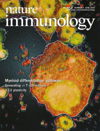
No. 6 June 2016
Nerlov and colleagues (p 666; News and Views by Sarrazin and Sieweke, p 609) use single-cell transcriptome profiling to investigate early differentiation into the hematopoietic lineage. Original image by Roy Drissen of cytospins of single pre–granulocyte-macrophage progenitor cells that express the transcription factor GATA-1 shows the simultaneous development of cells of the erythroid, megakaryocyte and eosinophil lineage. Artwork by Lewis Long
-
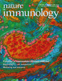
No. 5 May 2016
The physiological function of the mammalian chitinase AMCase is unclear. Wynn and colleagues (p 538) show that it is dispensable for allergic lung inflammation but is necessary for the clearance of intestinal helminths. The original image by Ian Moore and Kevin Bock shows a cross-section of a bronchiole from the lungs of a mouse exposed to helminth eggs. Artwork by Lewis Long.
-
No. 4 April 2016
Langerhans cells and tissue-resident memory CD8+ T cells require an active form of the cytokine TGF-β. Kaplan and colleagues (p 414) demonstrate that specific integrins expressed by epithelial cells activate latent TGF-β and that this is critical for maintaining resident immune cells in the skin and gut. The original image by Javed Mohammed shows epidermal whole mounts from mice doubly-deficient in the integrins β6 and β8 stained for the alloantigen Thy-1.1 (red) and MHC class II (green) after infection with LCMV. Artwork by Lewis Long.
-
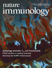
No. 3 March 2016
Regulatory T cells help mediate immunotolerance to self tissues. Chi and colleagues (p 277) find a crucial requirement for autophagy in orchestrating the suppressive activity of these cells in mice. The original image of hematoxylin-and-eosin–stained tissue by Peter Vogel shows extensive inflammation, fibrosis and infiltration of cells of the immune system in the esophagus. Artwork by Lewis Long.
-
No. 2 February 2016
Robbins and colleagues show that arteries are colonized by macrophages of various origins (p 159; News and Views p 117). The original image by Rickvinder Besla is a cross-section of the descending arch of the mouse aorta, stained for the chemokine CX3CL1 (red) and the adhesion molecule CD31 (yellow).Artwork by Lewis Long.
-
No. 1 January 2016
This month's Focus features a series of specially commissioned articles that discuss the most recent progress in understanding the ontogeny, functional diversity and activation plasticity of macrophages. See http://www.nature.com/ni/focus/macrophages/. Artwork by Lewis Long depicts élie Metschnikoff drawings of macrophages, as provided by S.H.E. Kaufmann from Metschnikoff, é. Immunität bei Infektionskrankheiten 1–456 (Verlag von Gustav Fischer, Jena, 1902).