Volume 10
-
No. 12 December 2003
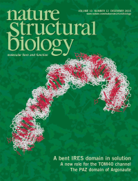
NMR structure ensemble of domain II of the HCV internal ribosome entry site (backbone white, bases red). At 25 kDa, domain II represents one of the largest pieces of RNA for which the structures have been determined by NMR. In solution, domain II adopts a distorted L-shape structure. This structure is similar to the overall shape of domain II bound to the 40S ribosome subunit. See pages 1033-1038.
-
No. 11 November 2003
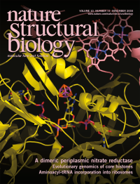
The structure of the heterodimeric periplasmic respiratory nitrate reductase (NapAB) from Rhodobacter sphaeroides showing the arrangement of the metallic cofactors. The diheme subunit NapB (pink) is associated with the catalytic NapA subunit (yellow), which contains both a 4Fe-4S cluster and a molybdenum cofactor. See pages 928–934.
-
No. 10 October 2003
The structure of tRNA guanine transglycosylase (TGT, gray surface and ribbon) from Zymomonas mobilis covalently linked to a stem-loop RNA substrate (green). TGT catalyzes the incorporation of a modified base in four tRNAs. The enzyme induces substantial conformation changes in the loop bases of the RNA substrate, and the structure of the covalent intermediate unambiguously identifies the aspartate residue that acts as the nucleophile for the reaction. See pages 781-788 and News and Views pages 772–773.
-
No. 9 September 2003
The three-dimensional structure of the bacteriophage T4 baseplate-tail tube complex based on cryo-EM reconstruction. The background image consists of electron microscope photographs of bacteriophage T4 stained with uranyl acetate or embedded in vitreous ice. See pages 688–693.
-
No. 8 August 2003
The crystal structure of the ear domain of GGA1 (red ribbon) in complex with a peptide (space-filling model, top) derived from p56. GGA1 is a monomeric adaptor protein for clathrin-coated vesicles; p56 is one of GGA1's protein ligand. This and the structure from a related study of the GGA3 ear domain reveal that conserved charged residues (purple space-filling model) on the GGA ear domains mediate the recognition of the hydrophobic phenylalanine in the peptides. Cover structure courtesy of B.M. Collins. See pages 599–606 and 607–613, News and Views pages 580–582.
-
No. 7 July 2003
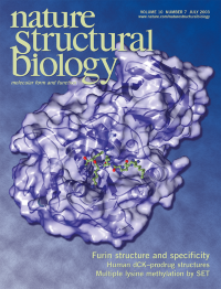
The crystal structure of a soluble mouse furin fragment (purple surface and ribbon) in complex with a peptide inhibitor (green and red model). Furin cleaves a wide range of proproteins, such as growth factors, and activates several bacterial toxins, including the anthrax toxin. The structure provides insights into the substrate specificity of furin. See pages 520-526.
-
No. 6 June 2003
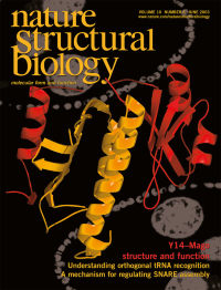
Crystal structure of the Drosophila protein Mago (yellow) in complex with Y14 (red). The complex is involved in post-splicing processes, such as nonsense-mediated mRNA decay and mRNA localization in Drosophila development. The background is a fluorescence microscope image showing co-localization of Y14 and Mago in the Drosophila egg chamber (Courtesy of O. Hachet and A. Ephrussi).
-
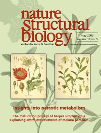
No. 5 May 2003
The human carboxylesterase 1 trimer (background, upper left) and the plants that generate two of this enzyme's substrates, heroin and cocaine. Chromolithography illustrations of the opium poppy Papaver sominferum (left) and the coca plant Erythroxylum coca (right) are reproduced from Franz Eugen Kohler's Medicinal Plants (Medizinal Pflanzen; 1883) and are reprinted here courtesy of the Missouri Botanical Garden, © 1995-2003 Missouri Botanical Garden http://ridgwaydb.mobot.org/mobot/rarebooks/
-
No. 4 April 2003
The overall structure of the U-box (ribbon and surface) from a yeast pre-mRNA splicing factor Prp19 resembles that of a RING finger domain. Two hydrogen-bonding networks (locations marked by green spheres) replace the Zn ions in the RING finger domain to stabilize the structure. The integrity of the U-box is important for its ubiquitin ligase activity and for the function of Prp19 in vivo. See pages 250–255.
-
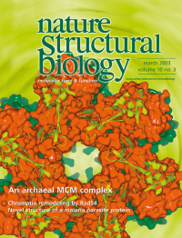
No. 3 March 2003
Crystal structure of the N-terminal region of the mini-chromosome maintenance (MCM) protein from Methanobacterium thermoautotrophicum. This region of the MCM protein forms a dodecameric complex containing two rings (orange and green surfaces) stacked back to back. The central channel is large enough to accommodate either double-or single-stranded DNA. Mutagenesis data support the role of the channel in DNA binding. See pages 160–167, and News and Views pages 148–150.
-
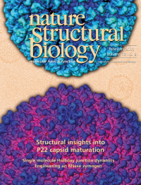
No. 2 February 2003
Cryo-EM structures of bacteriophage P22 at 9 å resolution. The overall fold and secondary structure elements of the capsid protein can be visualized in the structures of the procapsid (top) and the mature phage (bottom). Analysis provides insights into changes that occur during bacteriophage maturation. See pages 131–135.
-
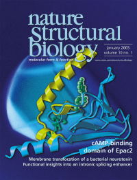
No. 1 January 2003
Structure of the ligand-free cAMP-binding domain of Epac2 (cyan) compared with the cAMP-bound form of protein kinase A (PKA, yellow; cAMP, transparent green). The Epac2 structure suggests a conserved mode of cAMP regulation for Epac2 and PKA (crucial residues are shown as ball-and-stick; green). Background is a single Epac2 crystal. From a design by G. Schulte. See pages 26–32.