Thank you for visiting nature.com. You are using a browser version with limited support for CSS. To obtain
the best experience, we recommend you use a more up to date browser (or turn off compatibility mode in
Internet Explorer). In the meantime, to ensure continued support, we are displaying the site without styles
and JavaScript.
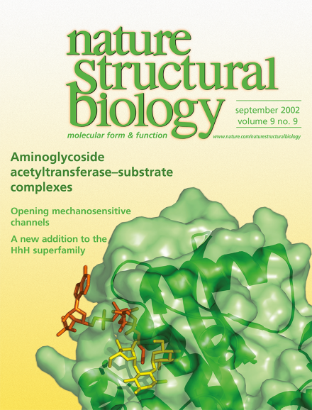
Mycobacterium tuberculosis amino-glycoside 2'-N-acetyltransferase (green surface and ribbons) in complex with cofactor coenzyme A (red) and antibiotic substrate ribostamycin (yellow). This enzyme can modify a broad range of aminoglycoside drugs and thereby confer antibiotic resistance to the bacteria. The structure provides insights into the modification reaction mechanism. It also suggests that the enzyme may participate in regulating the redox potential in mycobacterial cells. See pages 653–658.
Mechanosensitive ion channels from bacteria open like an iris, with the transmembrane helices tilting into the membrane to make a thin, wide structure with a 3 nm pore.
The structure of TAG, a DNA repair enzyme, reveals how evolutionary changes in the sequence of a conserved scaffold may enrich the mechanistic diversity of the DNA HhH glycosylase superfamily through a unique coupling of catalysis to substrate selectivity.
Molecular chaperones generally assist in the folding of proteins, and the cytoplasmic chaperone Hsp90, with its cofactors, additionally aids the activation of signaling proteins, including nuclear receptors. New evidence suggests that these chaperones also act to disassemble and down-regulate transcriptionally active nuclear receptor complexes.
A second three-dimensional crystal structure of the Ca2+-ATPase from the sarcoplasmic reticulum of rabbit muscle has been determined, this time in a Ca2+-free state. Its comparison with the previously determined Ca2+-bound state reveals details of structural changes at an atomic level in the catalytic cycle of a P-type ATP-dependent cation pump.