Thank you for visiting nature.com. You are using a browser version with limited support for CSS. To obtain
the best experience, we recommend you use a more up to date browser (or turn off compatibility mode in
Internet Explorer). In the meantime, to ensure continued support, we are displaying the site without styles
and JavaScript.
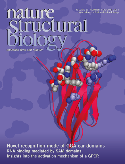
The crystal structure of the ear domain of GGA1 (red ribbon) in complex with a peptide (space-filling model, top) derived from p56. GGA1 is a monomeric adaptor protein for clathrin-coated vesicles; p56 is one of GGA1's protein ligand. This and the structure from a related study of the GGA3 ear domain reveal that conserved charged residues (purple space-filling model) on the GGA ear domains mediate the recognition of the hydrophobic phenylalanine in the peptides. Cover structure courtesy of B.M. Collins. See pages 599–606 and 607–613, News and Views pages 580–582.
Structural and functional analysis of WASP family cell signaling proteins shows that the same residues that are sequestered upon GTPase binding in the autoinhibited state are involved in binding to and activating Arp2/3 complex, WASP's downstream partner in the signaling cascade.
IgA triggers immune responses by binding to Fc receptors on cells of the immune system. The structure of IgA1-Fc in a complex with two molecules of FcαRI provides clues that may explain the selectivity of IgA-mediated immune events.
The structure of the calcium-bound core of human cardiac troponin reveals distinct subdomains connected by flexible linkers, implying that domain reorientation motions within the complex trigger muscle contraction.