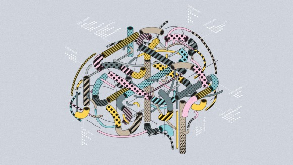
Thank you for visiting nature.com. You are using a browser version with limited support for CSS. To obtain
the best experience, we recommend you use a more up to date browser (or turn off compatibility mode in
Internet Explorer). In the meantime, to ensure continued support, we are displaying the site without styles
and JavaScript.
Four years ago, the NIH’s Brain Research through Advancing Innovative Neurotechnologies (BRAIN) Initiative Cell Census Network (BICCN) was launched, aiming to identify and catalog the diverse cells types in human, monkey and mouse brain. The first installment of this ambitious endeavor is now complete, with the comprehensive mapping of mammalian primary motor cortical cell type identities on a molecular level.
This collection features the reseach, datasets, methods and tools generated by this project. The flagship paper provides a comprehensive overview of the accomplishments while a variety of companion papers reveal the specifics of the data, the development of the tools and the application of the analytical tools. This dedicated collection also contains accompanying commentary in the form of an editorial, News and Views Forum and broad News Feature.
An atlas of the cell types found in the motor cortex of the brain has been built using various types of data. Two neuroscientists explain the technological feats involved in the project, as well as the utility of the resource for future research.
The BRAIN Initiative Cell Census Network has constructed a multimodal cell census and atlas of the mammalian primary motor cortex in a landmark effort towards understanding brain cell-type diversity, neural circuit organization and brain function.
An examination of motor cortex in humans, marmosets and mice reveals a generally conserved cellular makeup that is likely to extend to many mammalian species, but also differences in gene expression, DNA methylation and chromatin state that lead to species-dependent specializations.
A comprehensive survey of the epigenome from 45 regions of the mouse cortex, hippocampus, striatum, pallidum and olfactory areas using single-nucleus DNA methylation sequencing enables identification of 161 cell clusters with distinct locations and projection targets and provides insights into the regulatory landscape underlying neuronal diversity and spatial regulation.
As part of the BICCN consortium, the authors used a single-cell transcriptomic imaging method to produce a highly defined atlas of cell types across the mouse primary motor cortex.
Single-cell transcriptomic, morphological and electrophysiological characteristics are combined to classify more than 1,300 neurons from mouse motor cortex.
Combined patch clamp recording, biocytin staining and single-cell RNA-sequencing of human neurocortical neurons shows an expansion of glutamatergic neuron types relative to mouse that characterizes the greater complexity of the human neocortex.
Multi-modal analysis is used to generate a 3D atlas of the upper limb area of the mouse primary motor cortex, providing a framework for future studies of motor control circuitry.
Quantitative analysis of the methylation of mouse cortical neurons that project to different cortical and subcortical target regions provides insight into genetic mechanisms that contribute to differences in cell function.
Sparse labelling and whole-brain imaging are used to reconstruct and classify brain-wide complete morphologies of 1,741 individual neurons in the mouse brain, revealing a dependence on both brain region and transcriptomic profile.
A combination of genetic strategies and tools is used to define and fate-map different subtypes of glutamatergic pyramidal neurons according to their developmental and molecular programs, providing insight into the assembly of cortical processing networks.
RNA-sequencing analysis of the prenatal human brain at different stages of development shows that areal transcriptional signatures are dynamic and coexist with developmental and cell-type signatures.
Analysis of chromatin state at a single-cell level in samples of developing human forebrain demonstrate both cell-type-specific and region-specific changes during neurogenesis.
A single-cell atlas of the developing mouse cortex provides a temporal and spatial assessment of the molecular logic that drives the establishment and organization of cortical cell types.
Eze et al. use single-cell sequencing and immunohistochemical validation to create an atlas of early human brain development. In the telencephalon, they discover a diversity of progenitor subtypes, including two that are enriched in humans.
The superior colliculus (SC) receives diverse cortical inputs to drive many behaviors. Here, based on comprehensive mapping of cortico-tectal projections, the authors refined the superior colliculus into medial, centromedial, centrolateral, and lateral zones, and characterized the input-output connectivity and morphology of neurons in each zone that serve the role of SC in goal-directed behaviors.
Alternative RNA splicing varies across the brain. Its mapping at single cell resolution is unclear. Here, the authors provide a spatial and single-cell splicing atlas reporting brain region- and cell type-specific expression of different isoforms in the postnatal mouse brain.
Advances in large-scale connectivity mapping of the brain require efficient computational tools to detect fine structures across large volumes of images, which poses challenges. The authors introduce a hybrid architecture that incorporates topological priors of neuronal structures with deep learning models to improve semantic segmentation of neuroanatomical image data.
Single cell analysis of transposase-accessible chromatin is deepening our understanding on the origins of cellular diversity, yet methods are limited by data sparsity. Here, the authors introduce SnapATAC, a pipeline to resolve cellular heterogeneity and reveal candidate regulatory elements across different cell populations.
HD-fMOST is a microscopy technique for imaging large samples at high throughput and with high definition, which is achieved with a line-illumination modulation approach. The technology is illustrated by imaging fluorescently labeled neurons in whole mouse brains.
This protocol describes a scalable approach for joint profiling of chromatin accessibility and gene expression in single cells or nuclei that relies on multiple rounds of combinatorial indexing and can identify cell-specific regulatory elements.